EMG Diagnosis of Left Brachial Plexopathy: A Highly Skilled Diagnostic Procedure
1. Patient Presentation & Clinical History
A 28-year-old young male presented to our clinic following a road traffic accident that occurred four months prior. The patient complained of progressive weakness of the left upper limb with tingling and parasthesia diffusely distributed throughout the entire left upper limb. The most distressing symptom for the patient was the inability to abduct his arm, which significantly impaired his functional capacity and quality of life.
The patient reported progressive wasting of the muscles in his shoulder and upper arm, which he noticed over the weeks following the accident. He was unable to perform activities of daily living with his left arm and sought specialized neurological evaluation to determine the exact site and nature of the nerve injury.
- Road traffic accident 4 months prior
- Left upper limb weakness and wasting
- Diffuse tingling and parasthesia
- Inability to abduct arm (shoulder abduction loss)
- Loss of strength at shoulder, elbow, and wrist
- Minimal ulnar sensations preserved
2. Clinical Examination Findings
Detailed neurological examination revealed a precise pattern of weakness and preserved function that was critical for localization:
Motor Examination:
- Shoulder abduction: Completely lost (deltoid paralysis)
- Elbow function: Complete weakness at elbow flexion and extension
- Wrist function: Complete weakness at wrist flexion and extension
- Hand intrinsic muscles (C8-T1): Relatively spared; patient retained ability to abduct little finger
- Muscle atrophy: Significant wasting in shoulder girdle and arm musculature
Sensory Examination:
- Minimal ulnar sensations present in medial hand
- Reduced sensation in C5-C7 dermatomes
- Relatively preserved sensation in C8-T1 distribution (consistent with lower trunk involvement)
Clinical Significance: The sparing of C8-T1 innervated intrinsic hand muscles while the shoulder, elbow, and wrist were completely paralyzed strongly suggested involvement of the upper and middle trunks of the brachial plexus, with possible partial involvement of the lower trunk.
3. Anatomy of the Brachial Plexus: Understanding the Pathology
The brachial plexus is formed from nerve roots C5, C6, C7, C8, and T1. These roots organize into three trunks: upper (C5-C6), middle (C7), and lower (C8-T1). Each trunk then divides into anterior and posterior divisions, which further organize into three cords (lateral, medial, and posterior) before forming the terminal nerves.
| Brachial Plexus Component | Nerve Roots | Key Motor Functions |
|---|---|---|
| Upper Trunk | C5-C6 | Shoulder abduction (supraspinatus, infraspinatus), elbow flexion (biceps) |
| Middle Trunk | C7 | Elbow extension (triceps), wrist extension, finger extension |
| Lower Trunk | C8-T1 | Intrinsic hand muscles, finger abduction/adduction |
In this patient's case, the clinical pattern of weakness—loss of shoulder abduction (upper trunk C5-C6) and loss of wrist extension (middle trunk C7) with preserved finger abduction (lower trunk C8-T1)—pointed precisely to upper and middle trunk involvement.
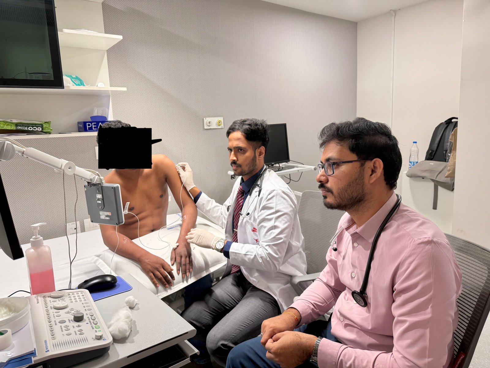
Dr. Rahul Gunde performing EMG procedure - demonstrating needle electrode placement technique for accurate nerve localization and trunk involvement assessment
4. The Highly Skilled EMG Diagnostic Procedure
Electromyography (EMG) is one of the most precise and specialized diagnostic tools in neurology. It requires extensive training, clinical expertise, and refined technical skill to perform accurately. In cases of brachial plexopathy, EMG is essential for localizing the exact site of nerve injury and determining the nature of the lesion (preganglionic vs. postganglionic).
Why EMG is a Highly Skilled Procedure:
- Precise needle placement: Requires intimate anatomical knowledge to position electrodes in specific muscles at exact depths
- Patient cooperation: Requires skilled communication to guide patient through various muscle activations
- Real-time interpretation: Demands ability to recognize normal vs. abnormal motor unit action potentials in real-time
- Pattern recognition: Requires expertise to identify specific patterns (denervation, reinnervation, myopathic patterns) and differentiate them
- Nerve localization: Demands knowledge of anatomical variations and skill in identifying subtle changes
- Multiple muscle sampling: Requires systematic testing of multiple muscles in different nerve distributions to map the lesion
EMG Methodology in This Case:
We performed a comprehensive EMG study with needle electrode examination of multiple muscles innervated by different branches of the brachial plexus to systematically map the extent and location of the nerve injury.
Muscles examined included:
- Supraspinatus (upper trunk via suprascapular nerve)
- Deltoid (upper trunk via axillary nerve)
- Biceps (upper trunk via musculocutaneous nerve)
- Triceps (middle trunk via radial nerve)
- Extensor carpi radialis (middle trunk via radial nerve)
- Wrist flexors (middle trunk)
- Abductor pollicis brevis (lower trunk via median nerve)
- First dorsal interosseous (lower trunk via ulnar nerve)
- Abductor digiti minimi (lower trunk via ulnar nerve)
5. EMG Findings & Lesion Localization
The EMG study revealed a precise pattern of denervation and motor unit abnormalities that allowed us to accurately localize the nerve injury:
Key EMG Findings:
- Upper trunk (C5-C6): Widespread denervation in supraspinatus, infraspinatus, deltoid, and biceps muscles
- Middle trunk (C7): Denervation in triceps, wrist extensors, and finger extensors
- Lower trunk (C8-T1): Partial involvement noted only in C8-C7 distribution of median nerve
- Spared functions: Intrinsic hand muscles (first dorsal interosseous, abductor digiti minimi) demonstrated normal motor unit recruitment, confirming partial preservation of lower trunk
Denervation Pattern Details:
- Fibrillation potentials: Present in upper and middle trunk-innervated muscles, indicating acute denervation
- Positive sharp waves: Observed in muscles with recent denervation
- Motor unit action potentials: Reduced recruitment in affected muscles; partially reduced in lower trunk muscles
- Reinnervation signs: Early polyphasia noted in some muscles, suggesting early reinnervation sprouting (4 months post-injury)
Critical Diagnostic Distinction: Postganglionic Lesion
A crucial finding was the absence of root-level lesions. In preganglionic injuries (nerve root avulsion), we would expect denervation of paraspinal muscles (innervated directly by the root prior to plexus formation). The normal EMG of paraspinal muscles in this patient confirmed a postganglionic lesion—meaning the nerve roots were intact, but the injury occurred within the brachial plexus itself.
- Postganglionic lesion (this case): Injury distal to nerve root; paraspinal muscles spared; potential for better recovery
- Preganglionic lesion: Nerve root avulsion; denervation of paraspinal muscles; poor prognosis for recovery
EMG procedure documentation - showing the clinical assessment phase where electrical activity of denervated muscles is recorded and analyzed for accurate lesion localization
6. Clinical Interpretation & Diagnostic Conclusion
The integration of clinical examination findings with EMG results provided a precise diagnosis: Left brachial plexus injury affecting the upper and middle trunks, with partial involvement of the lower trunk, secondary to road traffic accident four months prior. The lesion is postganglionic in nature.
The pattern of trunk involvement suggests an injury mechanism where the traumatic force primarily affected the upper and middle trunk regions—likely from forceful traction or direct trauma to the shoulder and upper arm region. The relative sparing of the lower trunk suggests it was partially protected or minimally affected by the trauma.
Significance of the Four-Month Timeline:
- Denervation changes are well-established, confirming the postganglionic nature of the injury
- Early reinnervation signs suggest spontaneous nerve recovery processes have begun
- This timeline is optimal for assessing the severity and predicting recovery potential
- Prognosis for recovery depends on the nature of the trunk injury (stretch vs. partial laceration)
7. Management & Prognosis
Treatment Approach:
Based on EMG findings confirming postganglionic injury with early reinnervation signs:
- Conservative management: Continue monitoring with serial EMG studies at 6-8 week intervals
- Physical therapy: Aggressive physiotherapy to prevent contractures and maintain passive range of motion
- Functional electrical stimulation: May be considered for denervated muscles to maintain muscle bulk
- Surgical consultation: If no improvement after 3-4 months or signs of complete transection, refer for microsurgical exploration and repair
Prognosis Indicators:
- Postganglionic lesion (better recovery potential than preganglionic)
- Early signs of reinnervation at 4 months
- Partial preservation of lower trunk function
- Young age (28 years) promotes neuroplasticity and recovery
8. Key Clinical Learnings & Prevention
Critical Learning Points:
- Pattern recognition: The preservation of C8-T1 functions while losing C5-C7 functions is pathognomonic for upper and middle trunk injury
- EMG's role: EMG transforms clinical suspicion into precise anatomical diagnosis and prognostic assessment
- Postganglionic vs. preganglionic: This distinction is crucial for prognosis and guides surgical decision-making
- Muscle sampling strategy: Systematic sampling across multiple nerves ensures accurate lesion localization
- Timeline importance: EMG timing (4 months post-injury) allows assessment of denervation acuity and reinnervation potential
Prevention of Brachial Plexus Injuries:
- Road safety: Use seat belts and avoid arm-out-window positioning during driving
- Workplace safety: Proper protective equipment and ergonomics in high-risk occupations
- Contact sports: Appropriate protective gear and technique to minimize shoulder trauma
- Birth-related injury prevention: Proper obstetric technique during difficult deliveries (for neonatal brachial plexopathy)
Red Flag Danger Signs Requiring Urgent Evaluation:
- Acute, severe paralysis following trauma (may indicate complete transection)
- Progressive weakness despite expected recovery timeline
- Signs of root avulsion (Horner's syndrome, diaphragmatic paralysis)
- Associated vascular compromise with cool, pale limb
- Severe pain (neuropathic pain may indicate poor prognosis)
9. Frequently Asked Questions About EMG and Brachial Plexopathy
EMG involves needle electrode insertion, which causes minimal discomfort similar to blood draw. Brief needle-related discomfort is expected, but the test provides invaluable diagnostic information. We use topical anesthesia and minimize patient discomfort through skilled technique.
A comprehensive brachial plexus EMG study typically takes 45-60 minutes, depending on the extent of involvement and number of muscles requiring examination.
Yes, EMG findings regarding denervation acuity, presence of reinnervation signs, and assessment of postganglionic vs. preganglionic injury help predict recovery potential. Serial EMG studies (at 6-8 week intervals) provide the most accurate prognostic information.
NCS measures electrical conduction along nerves and is useful for demyelinating disorders. EMG examines individual muscle electrical activity and is crucial for localizing nerve injuries, assessing denervation, and predicting prognosis. Both tests together provide comprehensive evaluation.
Not necessarily. EMG findings guide this decision. If signs of reinnervation are present and progressive improvement occurs, conservative management with physical therapy is preferred. Surgery is considered when EMG shows no improvement after 3-4 months or signs of complete nerve transection.
Ideally, EMG should be performed 3-4 weeks post-injury to allow denervation changes to develop. Earlier studies may miss denervation. Later studies (after 3-4 months) assess reinnervation potential and recovery trajectory.
Yes, with microsurgical nerve repair techniques. EMG findings help identify complete transections, and early surgical consultation (within 2-3 months) provides the best outcomes for functional recovery through end-to-end repair or grafting.
Recovery depends on injury type. Postganglionic injuries (like this case) have better prognosis: 50-70% achieve functional improvement. Young age, early intervention, and patient compliance with therapy improve outcomes significantly.
Have Similar Nerve Injury Symptoms?
If you or someone you know has experienced trauma to the shoulder or upper limb with persistent weakness and numbness, specialized neurological evaluation including EMG may be beneficial.
Contact Dr. Rahul Gunde for Expert Neurological Evaluation:
📍 KIMS Kompally, Hyderabad
📍 Lifespan Jeedimetla, Hyderabad